17TH WORLD CONGRESS ON CONTROVERSIES IN OBSTETRICS, GYNECOLOGY & INFERTILITY (COGI)
Our experience of laparoscopic myomectomy with temporary occlusion of internal iliac arteries
K. Puchkov1, N. Podzolkova2, Yu. Andreeva3, A. Dobychina1, V. Korennaya2
1Clinical and Experimental Surgery Center, Moscow, Russia; 2Medical Academy of Postgraduate Education, Moscow, Russia; 3Municipal Clinical Hospital N°̄ 67, Moscow, Russia
SUMMARY
In cases of “complicated” myomas in order to preserve the benefits of laparo- scopic access and to eliminate the shortcomings we developed a technique of lapa- roscopic myomectomy with temporary occlusion of internal iliac arteries. We apply the smooth vascular clamps «De Bakey» to the dissected arteries from both sides. This approach allows to keep control of the blood loss, to mobilize the myoma node without injuring the surrounding tissues, to perform the safe suture, that is extremely important for further fertility and pregnancy.
INTRODUCTION
In spite of the all advantages of laparoscopic access in cases of “complicated” myomas there are some problems connected with high risk of bleeding at the location site, probability of conversion, formation of a unreliable uterine scar un- der condition of bad wound visualization caused by continuing hemorrhage and consequently extensive use of electrosurgery in such conditions. We speak about «complicated» myoma in the following cases: size of node is more than 7-8cm, localization of node- interstitially with centripetal growth or on the posterior wall of uterus or in the rib area of the uterus. In these cases in order to eliminate the shortcomings and to preserve the benefits of laparoscopic access our clinic de- veloped a technique of laparoscopic myomectomy with temporary occlusion of internal iliac arteries.
MATERIAL AND METHODS
We have been successfully using this method since 2008. The parietal peritoneum is laparoscopically excised above the iliac arteries.
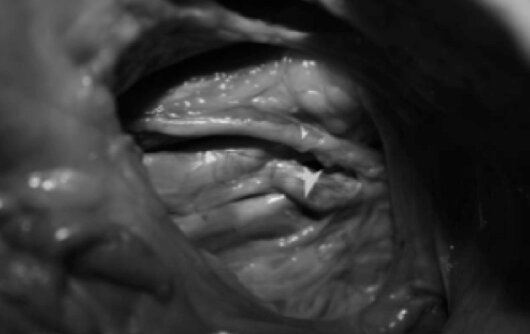
Photo 1 - Anatomy of ureter and iliac vessels (cadaver). Marked ureter and internal iliac artery.
Smooth vascular clamps “De Bakey” are introduced into the abdominal cavity by “Endoclinch” forceps. The clamps are applied on the dissected arteries from both sides.
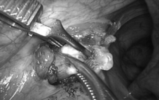
Photo 2 - Key step of the access: internal iliac artery occlusion with soft vascular forceps «De Bakey».
The incision of the uterus wall above the myoma node is performed by ultrasonic scissors (Auto Sonix Covidien) or using monopolar coagulation. The myoma node is extracted from the surrounding tissue by two 10mm forceps. We have never ob- served wound bleeding, so we have excellent opportunity to visualize the border of node. In these circumstances (without bleeding, with minimal electrosurgical dam- age of myometrium) we separate the node without the risk of uterine cavity opening.
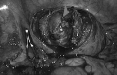
Photo. 3 - The myoma node is removed and endometrium border is good visualized.
While the node is being pulled out, we inject oxitocin intravenous ; more contracting uterus “pushes” the node helping it to get out, the wound surface is getting smaller. Un- der the conditions of good visualization we suture the wound in several layers making it safe and reliable. We use synthetic absorbable suture material for closing the wound. Convenient exposition is created by means of uterus manipulator. Myoma nodes are removed by morcellation. The uterus body is covered with antiadhesion barrier.
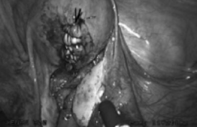
Photo. 4 - The uterus wound is sutured and covered with anti-adhesive barrier.
At the end of the operation the soft clamps are taken off and the blood flow in the iliac arteries is fully restored.
RESULTS
In the four-year period 204 (average 22-48y.o.) patients have been treated by the described method. 140 (68%) patients have had multiple “difficult” myomas (50-80 cm2 of volume and/or “inconveniently” located). The operative time (mean ± SD) was 65± 20 min. Time, spent on clamp placement, was 15± 5 min. Blood loss didn`t overcome 50 ml and mean was 40±10 ml. Patients stayed at hospital for 2-3 days. Average recovery period – 14 days. The early therapeutic outcomes showed shorter recovery period, less amount of analgetics needed and no infectious or thrombotic complications. No blood transfusion was needed. At the 4-year follow-up low recur- rence rates and good symptom control were obtained. Scar formation was controlled by the ultrasonic examination in 7, 30, 60 and 180 days after surgery The quality of the uterus scar we check by the dynamic sonographic control. We have had good quality of scar without hematoma in any cases.
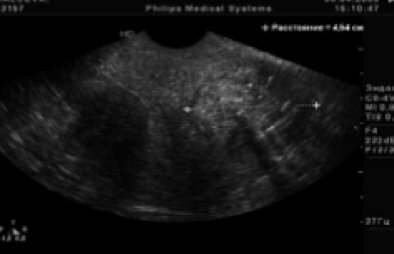
Photo. 5 - The sonographic picture after 7 days postoperation period.
Trough out this period we have had one complication (iliac vein injury) with no conversion to laparotomy.
CONCLUSIONS
This method is acceptable and feasible, as far as it has the following benefits for doctor and patients:
– provides control of the blood loss – doesn`t prolong the operation time – allows to apply laparoscopy even in difficult cases.

